The AI Companion to Your TPS

ART-Plan™

Our lighthouse applications focus on precision radiotherapy eliminating the gap from imaging to treatment and adaptation. Modular, scalable, and secure, our technology fully integrates within your clinical workflow.

Quality

Reliable, high-quality results derived from robust models created with data from leading global centers ensuring accuracy, confidence and physician acceptance

Simplicity

Automation seamlessly integrated within TPS workflows to accelerate job completion in the background without user supervision

Versatility

Works with any TPS to reduce time and complexity across the spectrum - from segmentation to planning to adaptive - for most indications.

Trusted Results.
Integrated Workflows.
End-to-End Coverage.

ART-Plan™

Play Video

Accelerate Segmentation

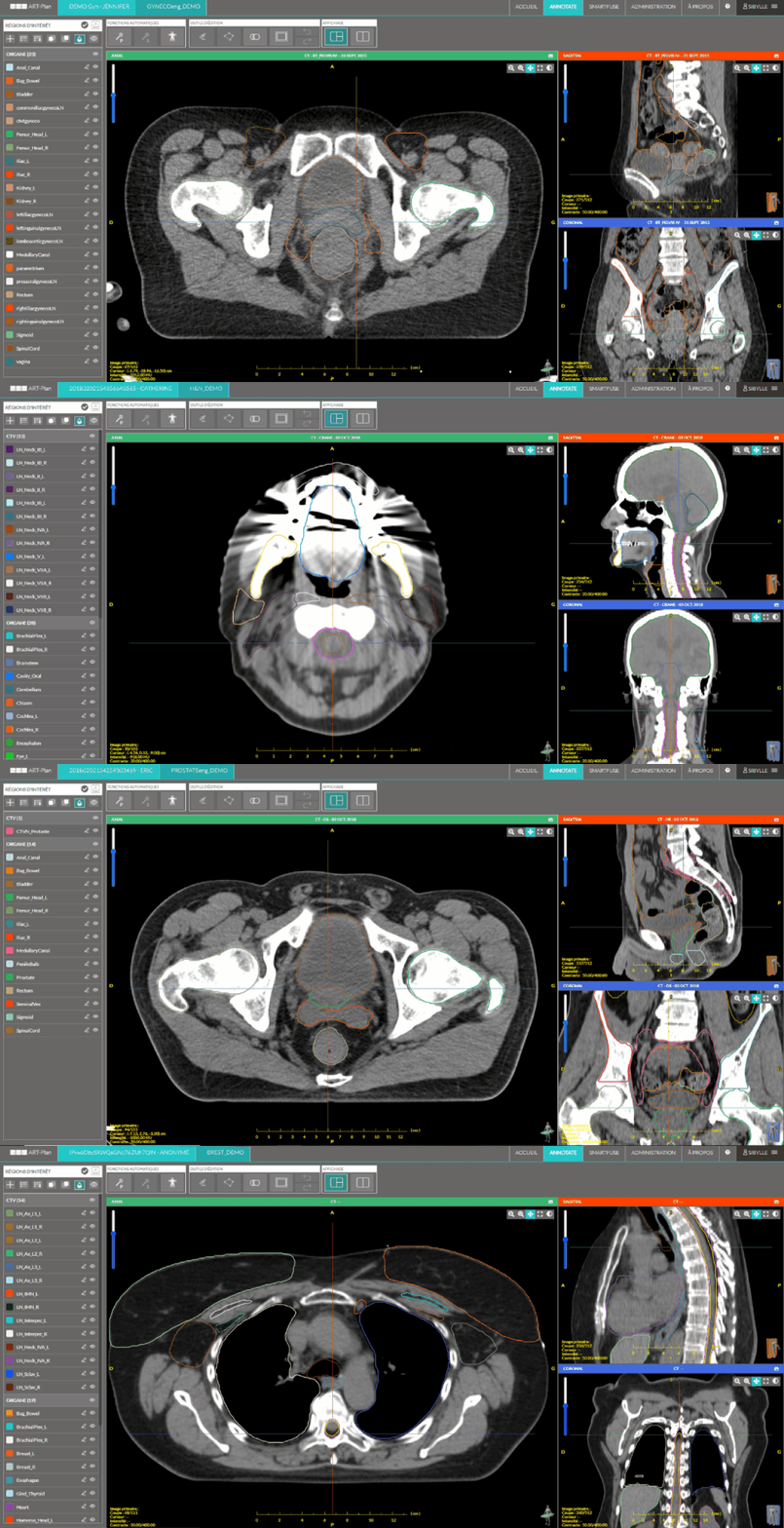
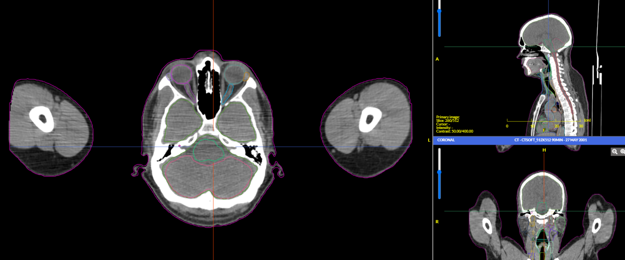
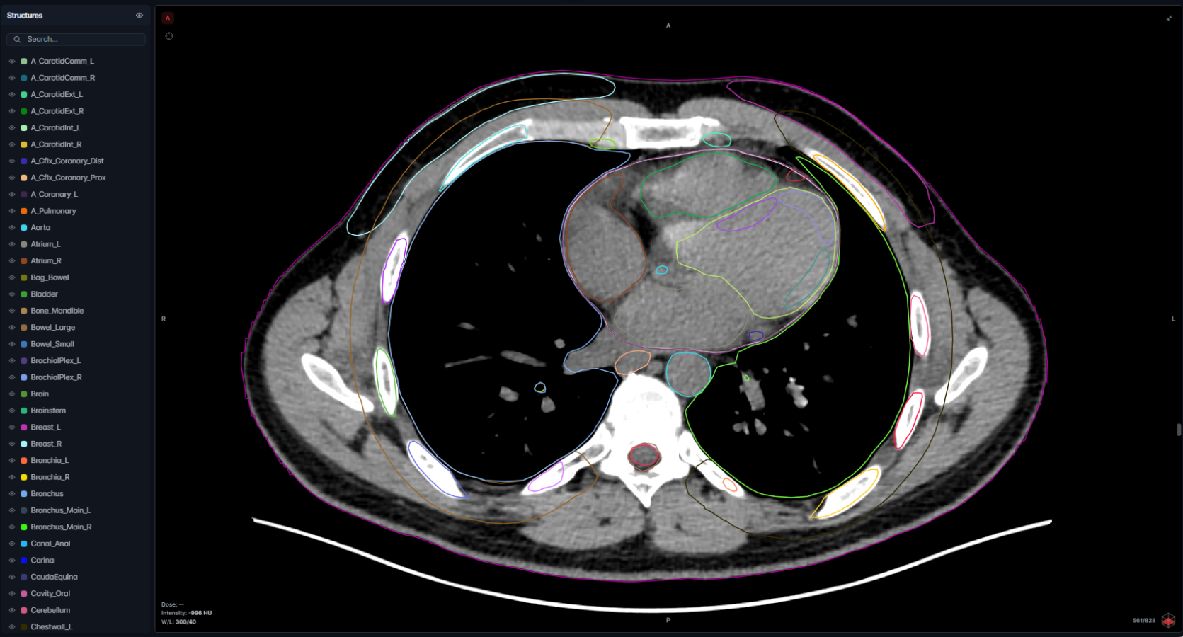
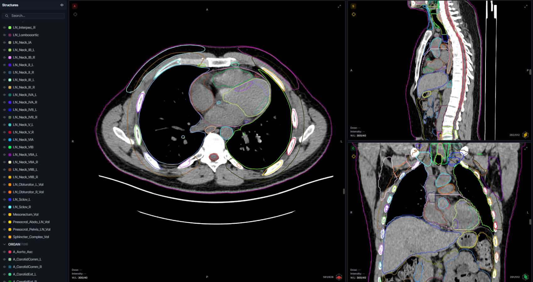
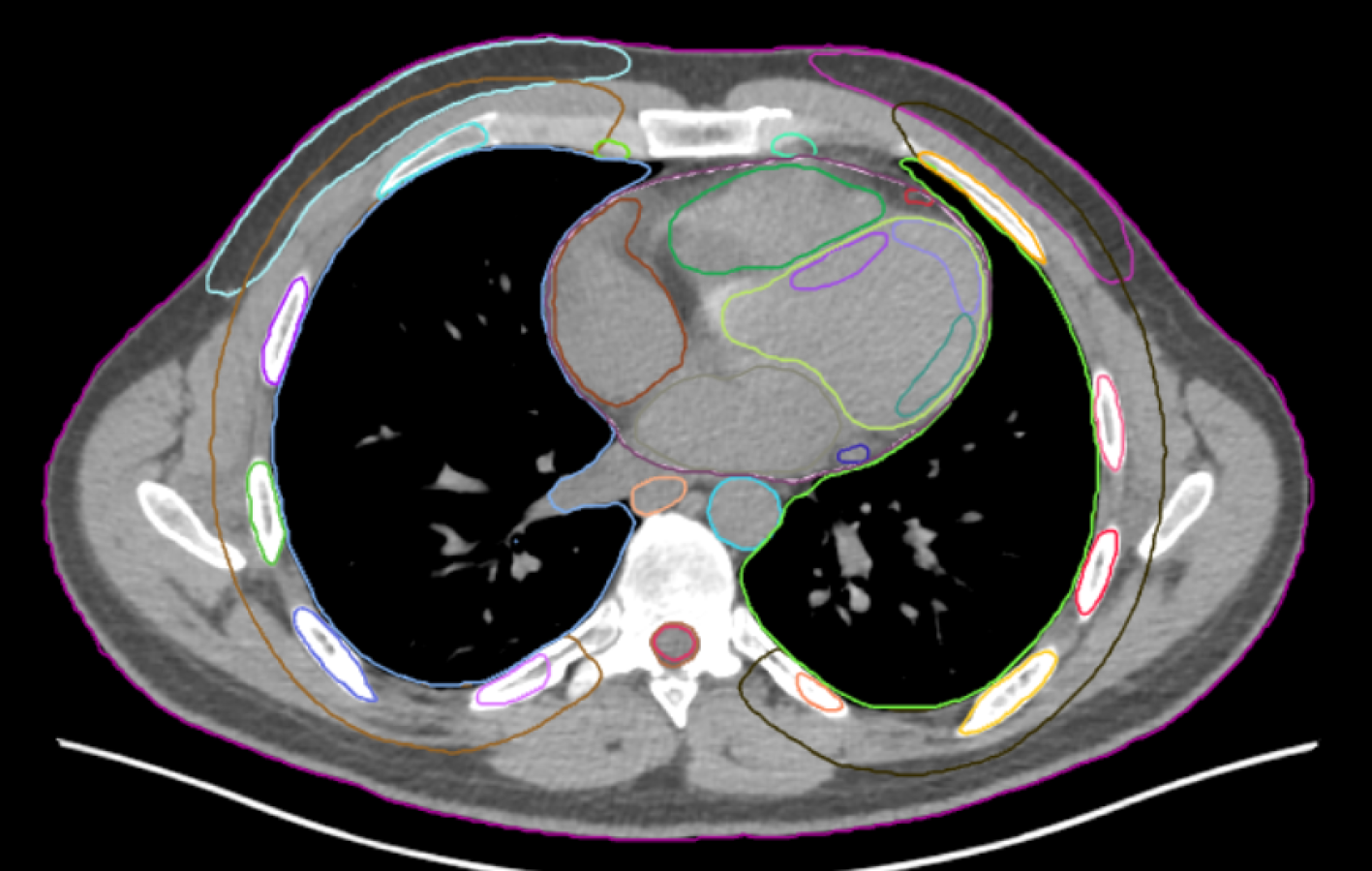
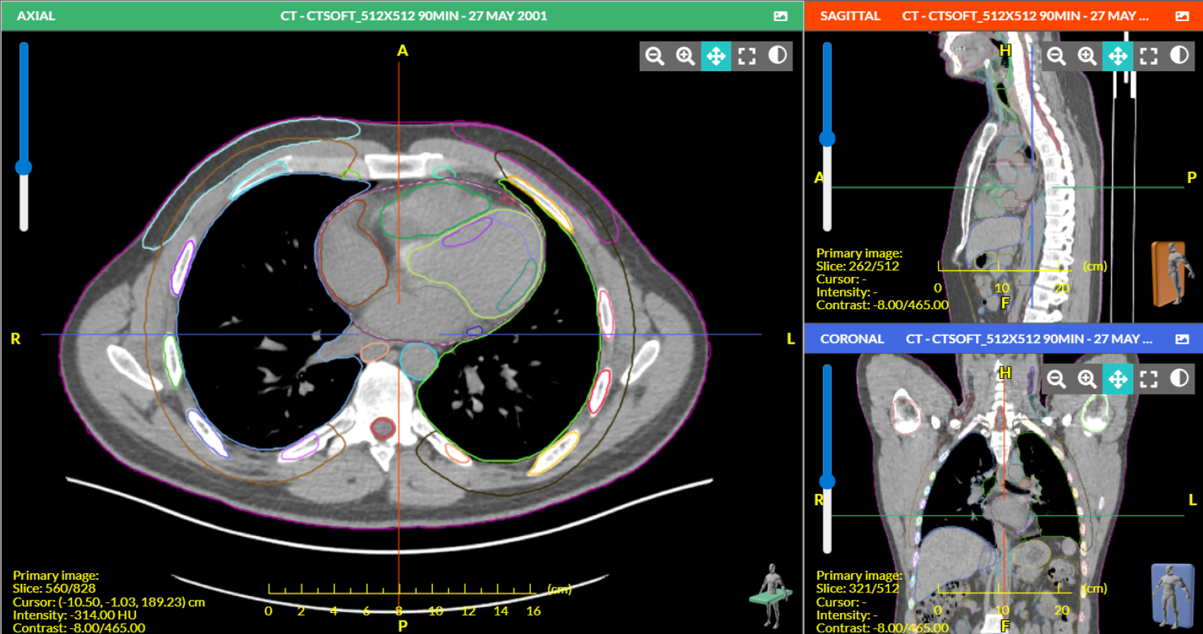
ART-Plan is an all-in-one solution for AI-based automatic delineation in minutes with the accuracy of clinical experts. Operating autonomously inside your current TPS, ART-Plan simplifies OAR and tumor segmentation with zero clicks to reduce manual tasks by >93%*.

* Doolan PJ et al. Frontiers in Oncology, 2023

Right the First Time

Our models are created with data from multiple leading academic centers, treating patients from diverse populations for a wide range of indications.

Physician Acceptance

All our models have been meticulously and continuously evaluated to ensure each organ and lymph node model is clinically acceptable.

Efficient

Expert-level RT plans in just a few minutes - eliminating manual variability and accelerating planning workflows.

Reduce Manual Tasks by >93%.
Accelerate Segmentation with Zero Clicks.

Segmentation

Key Features

OAR Segmentation

270+ OARs and LNs in minutes
Models for CT/MR/sCT

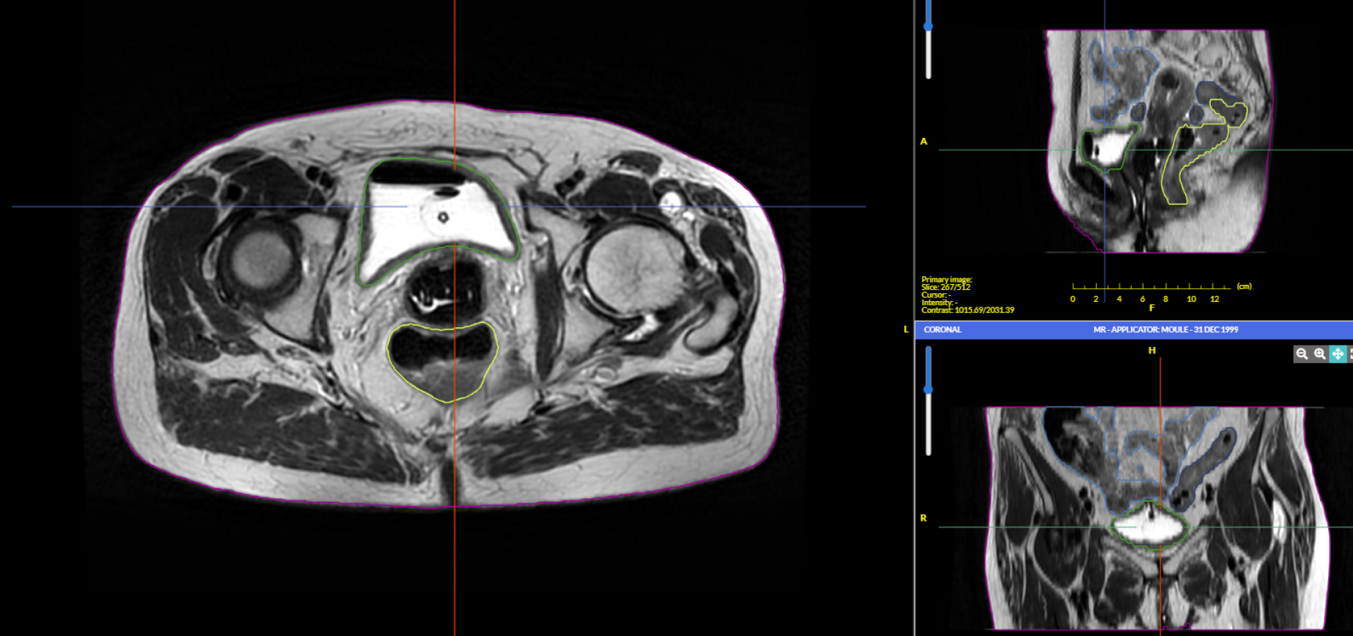
Brachytherapy Segmentation

MR OARs for brachytherapy
Bladder, rectum, sigmoid colon, small intestine

Tumor Segmentation

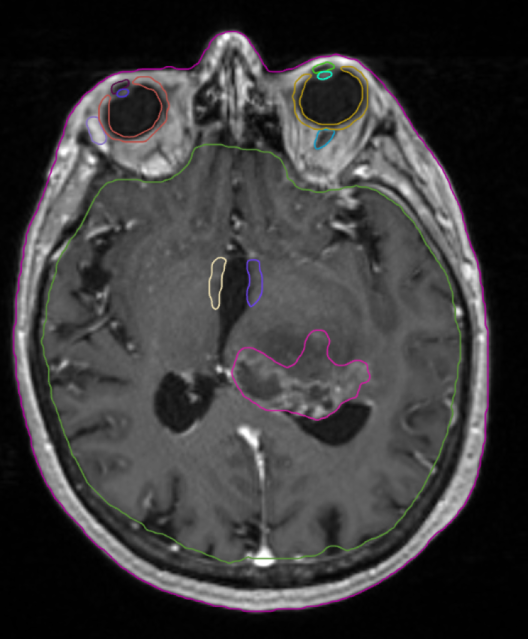
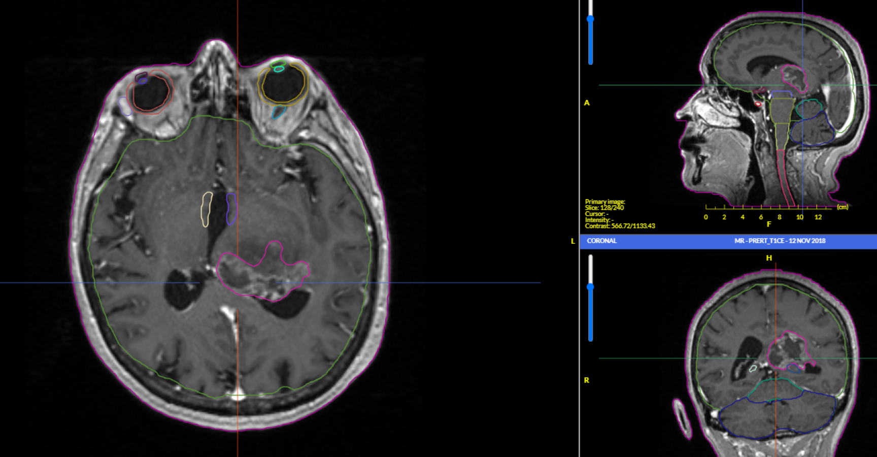
MR GBM and Multi-Mets

Fusion

Multi-modality, rigid and DIR
CT, MR, PET, CBCT + ITV from 4DCT

*Note that not all models are available in all markets

Segmentation Models

Structures
Library

OARs - Guidelines: DAHANCA 2020, Eekers et al 2021 (EPTN) and Mir et al 2020 (International consensus).

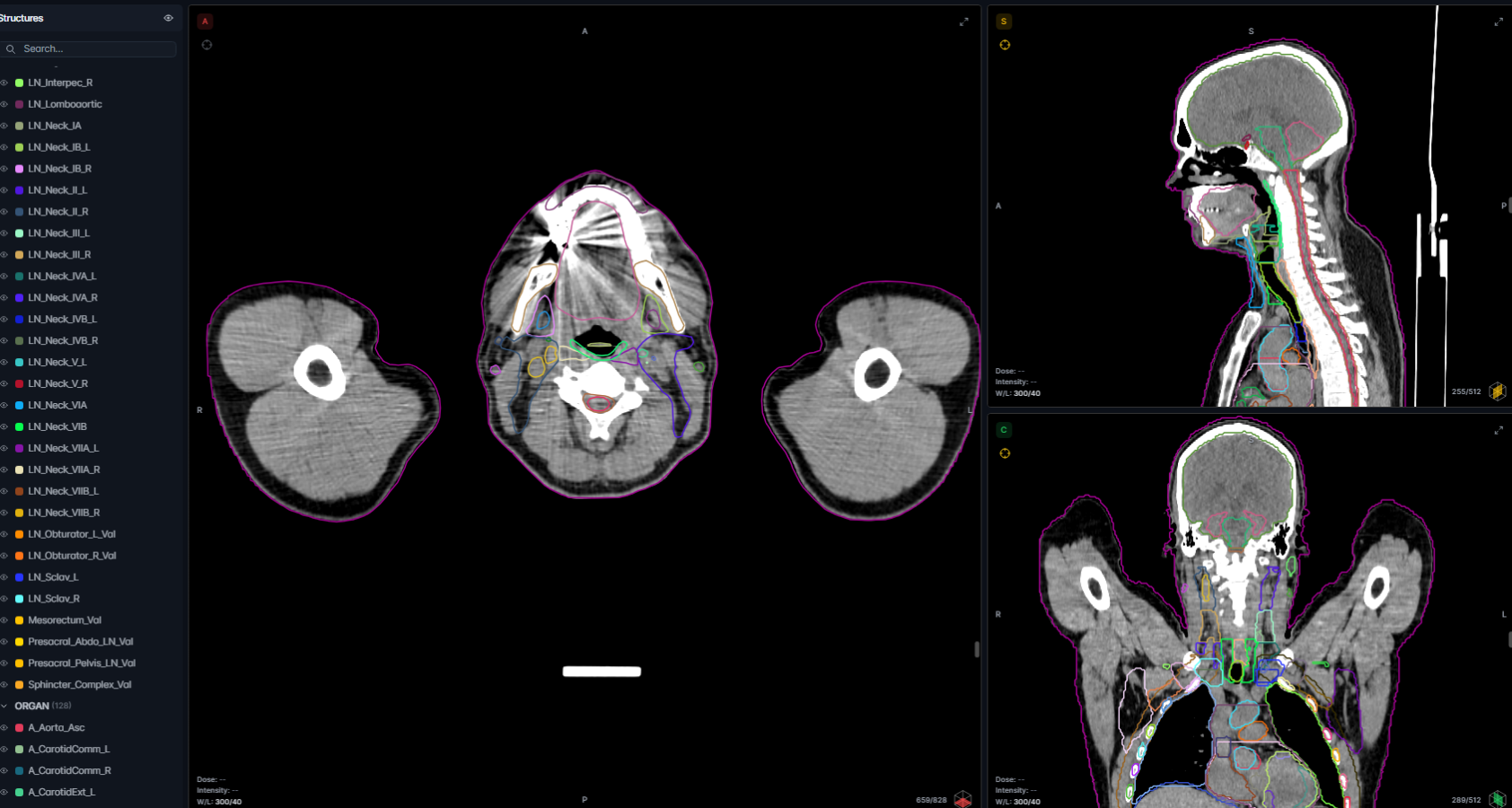
Brachial_Plex_(L/R), Brain, Brainstem, Cerebellum, Cochlea_(L/R), Common carotid artery (L/R), Esophagus, External carotid artery (L/R), External contours, External jugular vein(L/R), Eyeball_(L/R), Lens_Eye_(L/R), Glottic_Larynx, Internal carotid artery (L/R), Internal jugular vein (L/R), Lacrim_gland_(L/R), Larynx, Lips, Mandible, Mouth, Optic_chiasm, Optic_nerve_(L/R), Parotid_(L/R), Pharynx_Const_muscle, Pituitary_Gland

Spinal_Canal, Spinal_Cord, Submand_Gland_ (L/R), Supraglottic_Larynx, Thyroid_Gland, TM_Joint_(L/R), Trachea

LNs - Guidelines: Grégoire et al 2014,

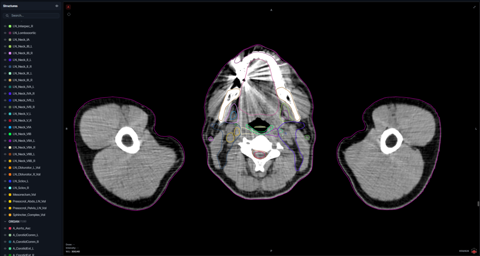
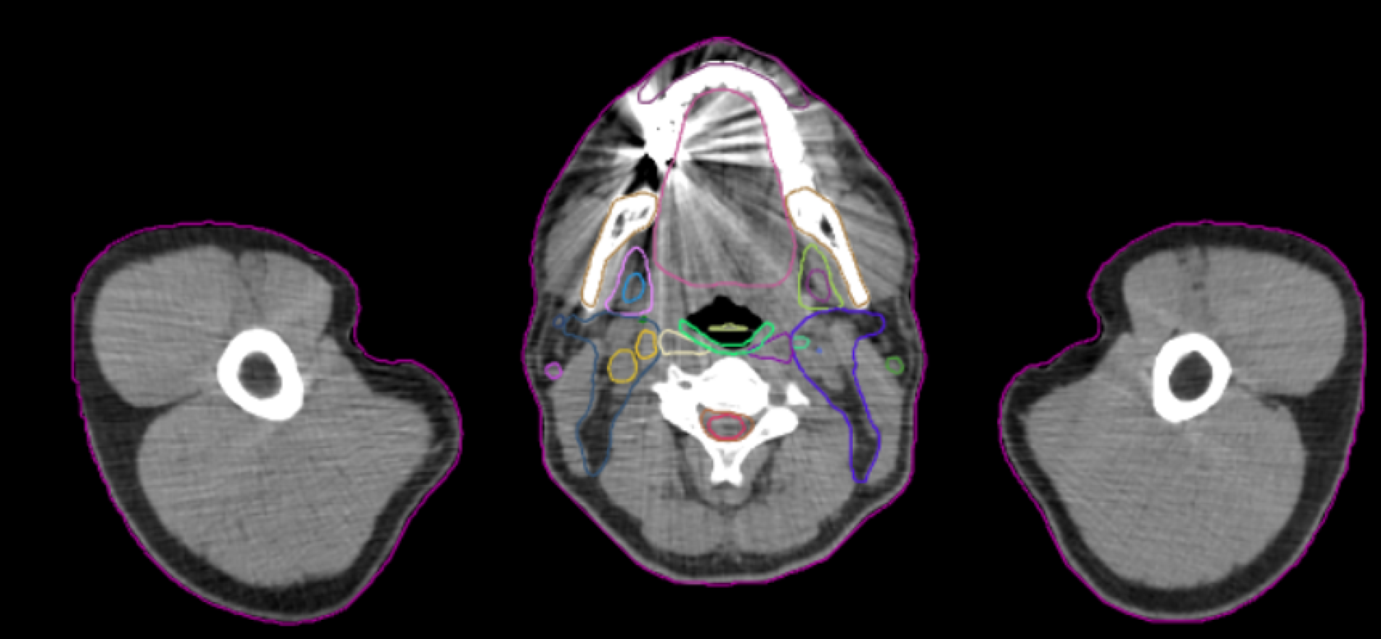
LN_Neck_IA, LN_Neck_IB_(L/R), LN_Neck_II_(L/R), LN_Neck_III_(L/R), LN_Neck_IVA_(L/R), LN_Neck_IVB_(L/R), LN_Neck_V_(L/R), LN_Neck_VIA, LN_Neck_VIB, LN_Neck_VIIA_(L/R), LN_Neck_VIIB_(L/R)

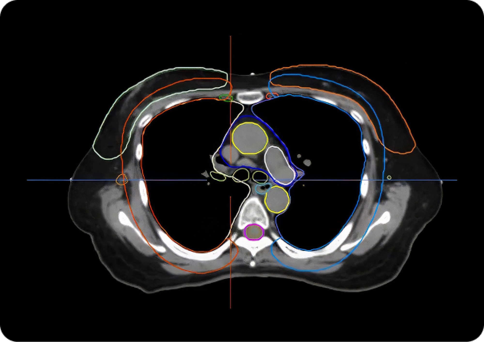
Includes SBRT lung and Heart substructures 

OARs – Guidelines: Kong 2011 (RTOG), Jabbour et al 2014 (RTOG), UK SABR Consortium 2019, De Rose et al, 2017, Duane et al 2017, Milo et al 2020, Mir et al 2020 

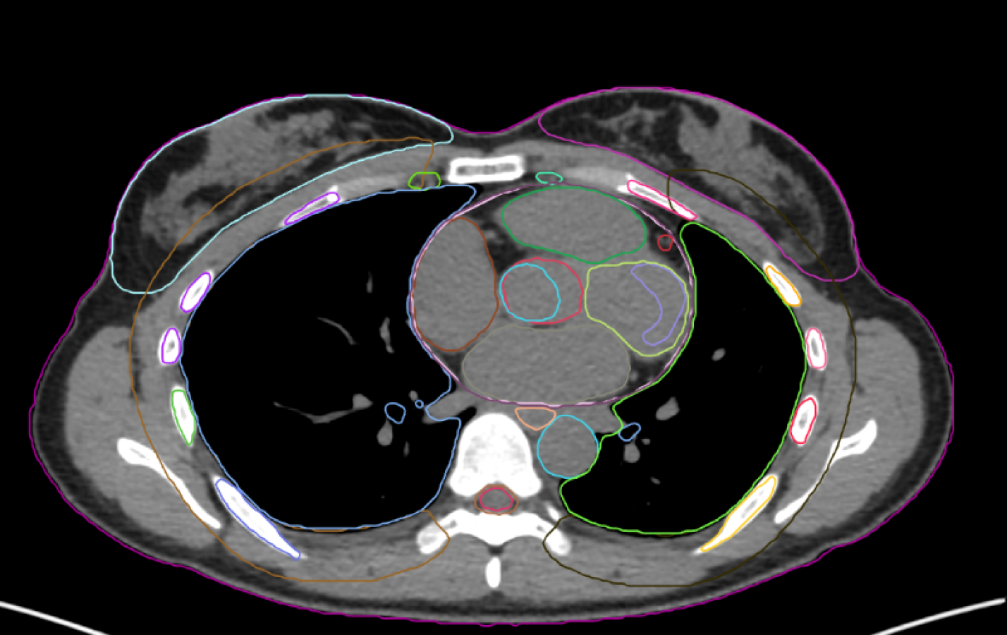
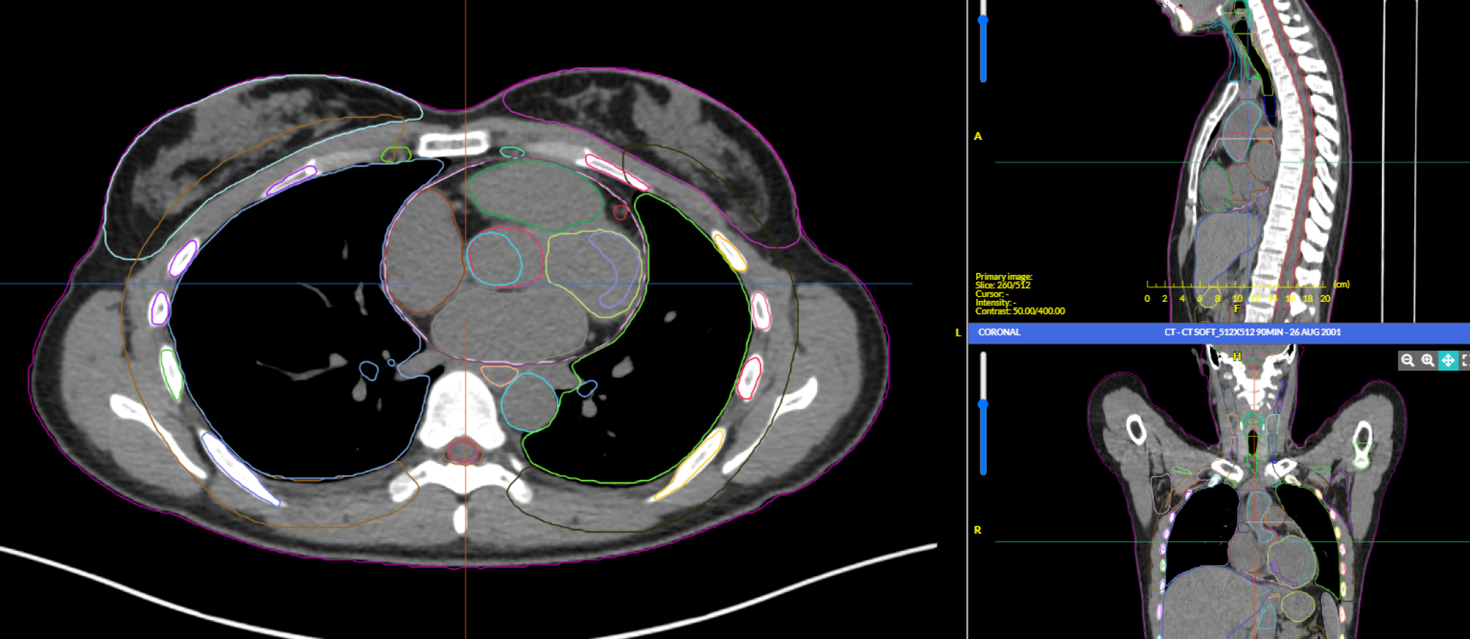
Aorta, Atrium_(L/R), Brachial_Plex_(L/R), Breast_(L/R), Bronchia_(L/R), Bronchial_Tree, Bronchus_(L/R), Cardiac_Aorta, Carina, Cauda Equina, Chestwall_(L/R), Coronary_Sinus, Distal_Circumflex_Coronary, Esophagus, Heart, Humerus_Head_(L/R), Kidney_(L/R), LAD_Coronary, L_Main_Coro_Art, Liver, Lung_(L/R), Pericardium, Proximal_Circumflex_Coronary, Pulmonary_Arter, Ribs 1-12_(L/R), Spinal_Canal, Spinal_Cord, Spleen, Stomach, Supraglottic_Larynx, Thyroid_Gland, Trachea, Vena_Cava_Inf, Vena_Cava_Sup, Ventri_(L/R), Ventri_L_Ant, Ventri_L_Api, Ventri_L_Inf, Ventri_L_Lat, Ventri_L_Sept,  

LNs - ESTRO - LN according to Offersen et al 2015 (ESTRO) 

LN_IMN_(L/R), LN_Interpec_(L/R), LN_Ax_L1_(L/R), LN_Ax_L2_(L/R), LN_Ax_L3_(L/R), LN_Ax_L4_(L/R) 

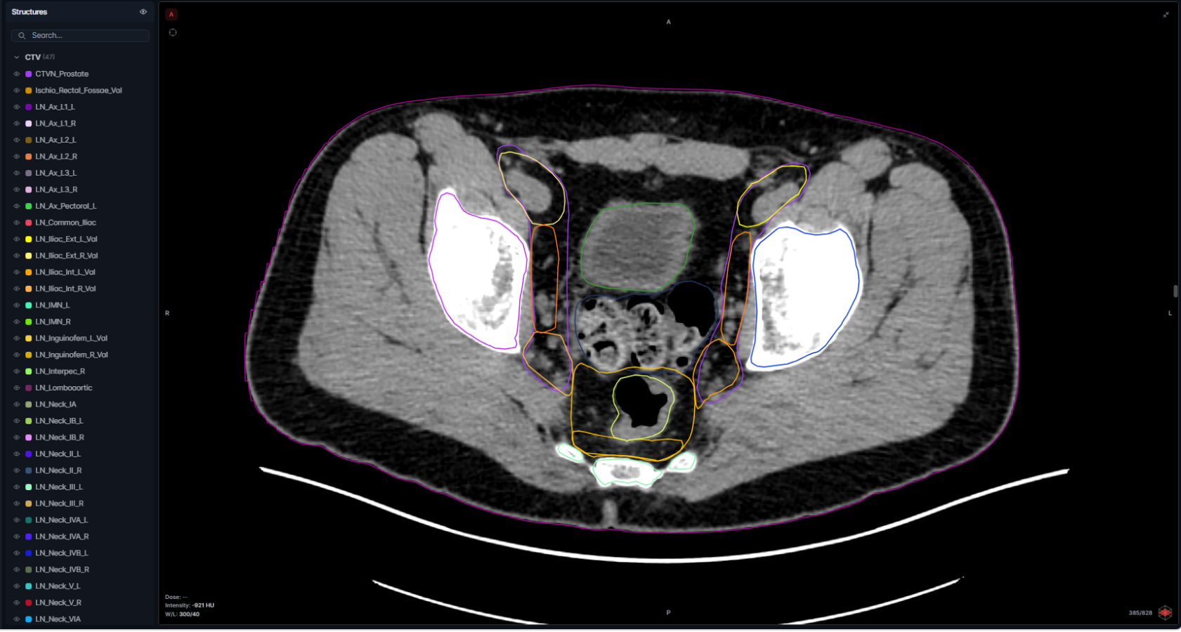
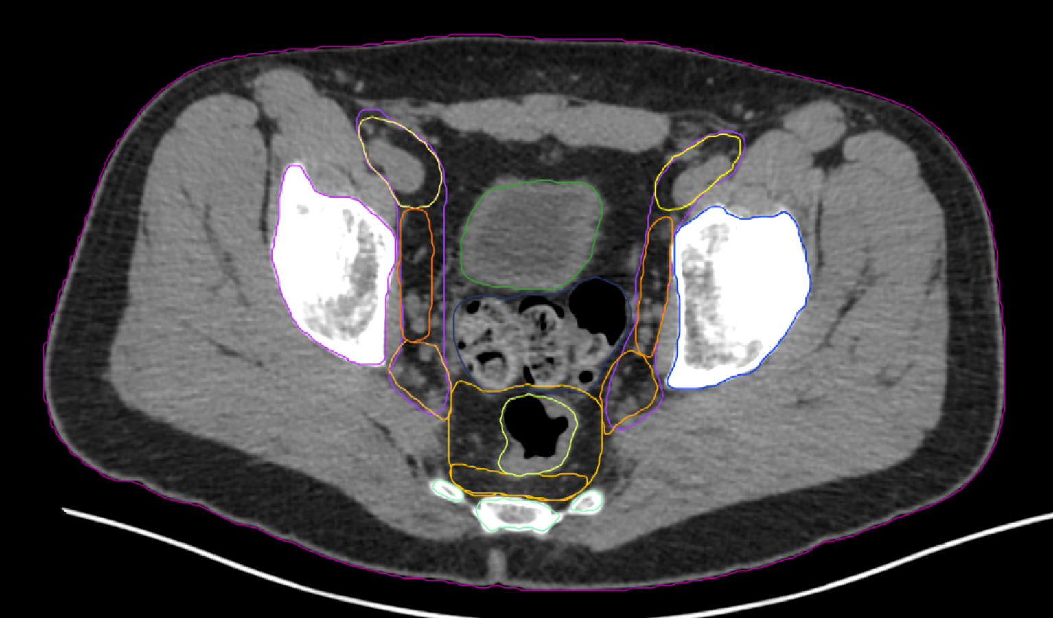
OARs - Guidelines: Gay et al 2012 (RTOG), Mir et al 2020, CTVN_Prostate as described in Hall et al 2021 

Anal_Canal, Bladder, Bowel_Bag, Cauda Equina, Duodenum, External contours, Femur_Head_(L/R), Illium_(L/R), Large_Bowel, Penile_Bulb, Rectum, Sacrum, Sigmoid_Colon, Small_Bowel, Spinal_Cord, VB_L4, VB_L5 

LNs - Guidelines: Valentini et al 2016 

Ischio_Rectal_Fossae_Val, LN_Iliac_Ext_L_Val, LN_Inguinofem_L_Val, LN_Iliac_Int_L_Val, LN_Obturator_L_Val, Mesorectum_Val, Presacral_Abdo_LN_Val, Presacral_Pelvis_LN_Val, LN_Iliac_Ext_R_Val, LN_Inguinofem_R_Val, LN_Iliac_Int_R_Val, LN_Obturator_R_Val, Sphincter_Complex_Val, LN_Common_Iliac, LN_Lomboaortic 

Regions of Interest 

CTVN_Prostate, Prostate, Seminal_Vesicle 

OAR - Guidelines: Gay et al 2012, Mir et al 2020 

Anal_Canal, Bladder, Bowel_Bag, Cauda Equina, Duodenum, External contours, Femur_Head_(L/R), Ilium_(L/R), Large_Bowel, Rectum, Sacrum, Sigmoid_Colon, Small_Bowel, Spinal_Cord, Vagina, VB_L4, VB_L5,  

LNs - Guidelines: Valentini et al 2016 

LN_Common_Iliac, LN_Iliac_(L/R), LN_Ingui_Gyn_(L/R), LN_Lomboaortic, LN_Presacral, Ischio_Rectal_Fossae_Val, LN_Iliac_Ext_L_Val, LN_Inguinofem_L_Val, LN_Iliac_Int_L_Val, LN_Obturator_L_Val, Mesorectum_Val, Presacral_Abdo_LN_Val, Presacral_Pelvis_LN_Val, LN_Iliac_Ext_R_Val, LN_Inguinofem_R_Val, LN_Iliac_Int_R_Val, LN_Obturator_R_Val, Sphincter_Complex_Val 

Region of Interest 

Parametrium, Utero_Cervix 

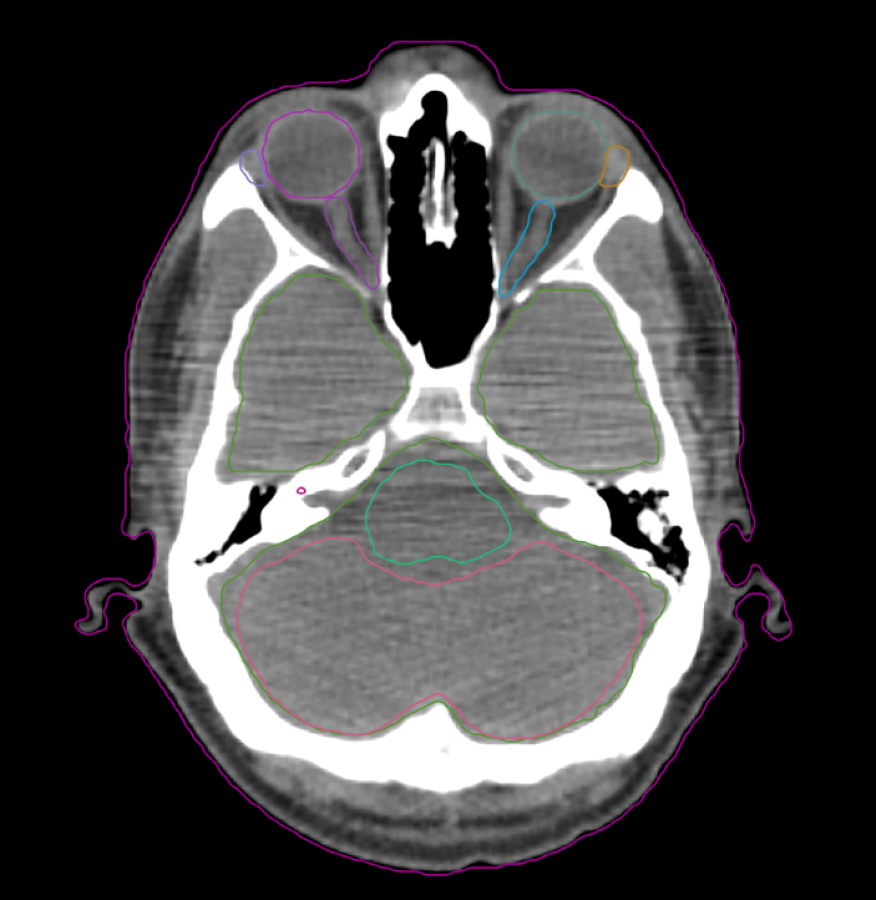
OAR - Guideline: Eekers et al 2021 (EPTN) 

Brain, Cerebellum_Ant, Cerebellum_Post, Cochlea_(L/R), Cornea_(L/R), Hippocampus_(L/R), Hypothalamus_(L/R), Lacrim_Gland_(L/R), Lens_Eye(L/R), Medulla_Oblong, Midbrain, Opt_Chiasm, Optic_nerve (L/R), Pituitary_Gland, Pons, Retina_(L/R), Spinal_Cord, VSCC_(L/R) 
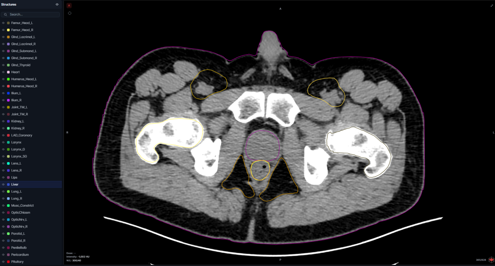
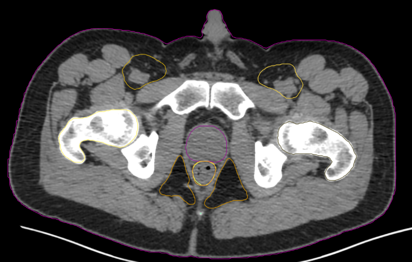
OARs - Guideline: Mir et al 2020 

Anal_Canal, Bladder, Bony_Pelvis_(L/R), Femur_Head_(L/R), Penile_Bulb, Rectum, Sacrum, Colon_Sigmoid, Small Bowel 

Regions of Interest  

Prostate, Seminal_Vesicle 

Pelvis OARs - Guideline: Mir et al 2020 

Anal_Canal, Bladder, Femur_Head_(L/R), Penile_Bulb, Rectum, Sigmoid_Colon,  

Abdo OARs - Guideline: Mir et al 2020 

Aorta_Abdomen, Duodenum, Kidney_(L/R), Large_Bowel, Liver, Pancreas, Stomach, Vena Cava_In 

Regions of Interest 

Prostate, Seminal_Vesicle 

OARs - Guidelines: EMBRACE studies, Pötter et al. 2018 (ESTRO) 

Bladder, Rectum, Sigmoid_Colon, Small_bowel 

  • Brain

    Reliable tumor and OAR segmentation for confident planning

  • Brain

    Reliable tumor and OAR segmentation for confident planning

  • Brain

    Reliable tumor and OAR segmentation for confident planning

  • Brain

    Reliable tumor and OAR segmentation for confident planning

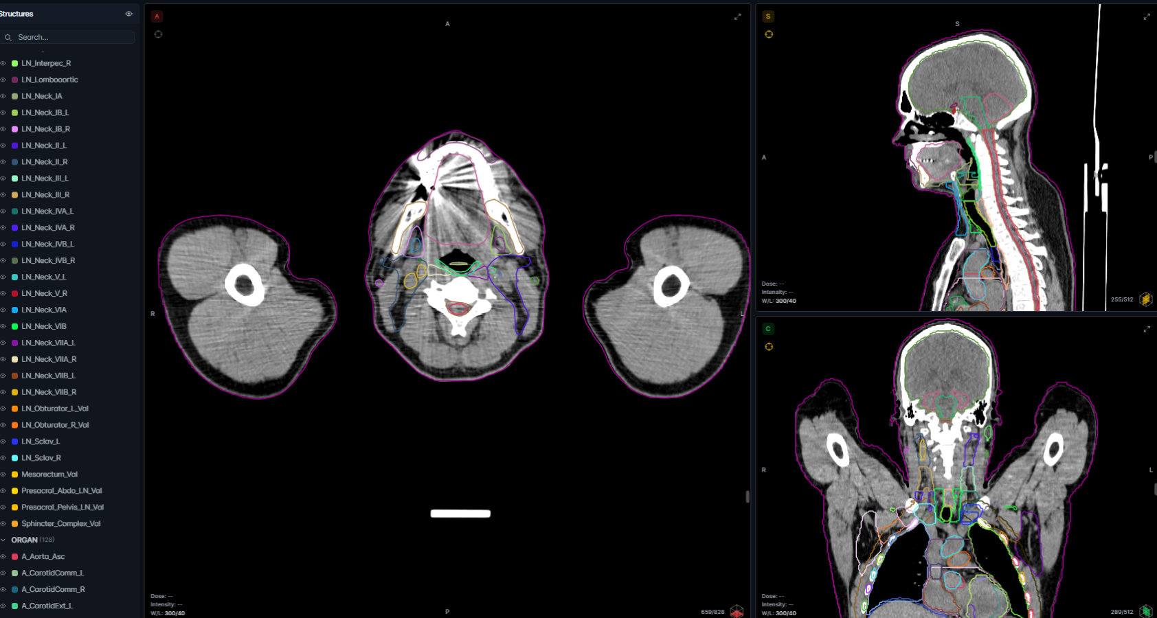
  • Head & Neck

    Precise OAR delineation in anatomically complex regions

  • Head & Neck

    Precise OAR delineation in anatomically complex regions

  • Head & Neck

    Precise OAR delineation in anatomically complex regions

  • Head & Neck

    Precise OAR delineation in anatomically complex regions

  • Breast

    Streamlined contouring with consistent adaptive workflows

  • Breast

    Streamlined contouring with consistent adaptive workflows

  • Lung & Thorax

    Robust segmentation supporting reliable planning in motion

  • Lung & Thorax

    Robust segmentation supporting reliable planning in motion

  • Lung & Thorax

    Robust segmentation supporting reliable planning in motion

  • Lung & Thorax

    Robust segmentation supporting reliable planning in motion

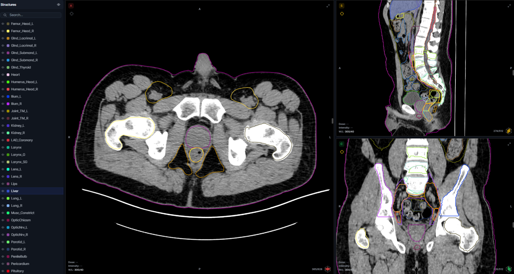
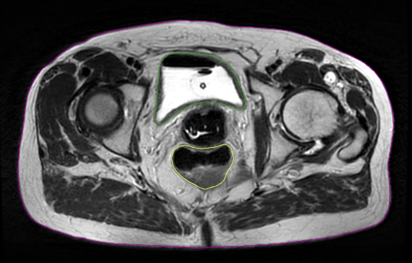
  • Male Pelvis

    Automated planning with integrated contouring and adaptation

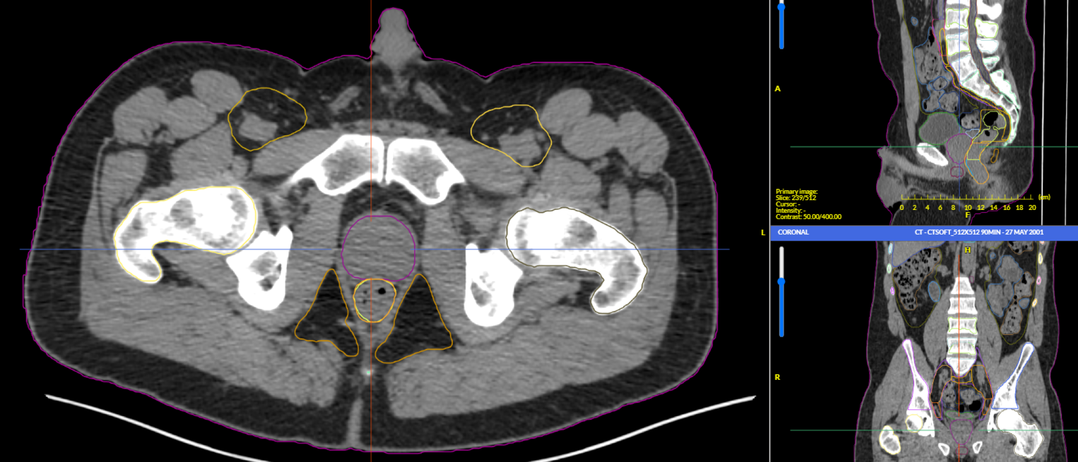
  • Male Pelvis

    Automated planning with integrated contouring and adaptation

  • Male Pelvis

    Automated planning with integrated contouring and adaptation

  • Male Pelvis

    Automated planning with integrated contouring and adaptation

  • Male Pelvis

    Automated planning with integrated contouring and adaptation

  • Male Pelvis

    Automated planning with integrated contouring and adaptation

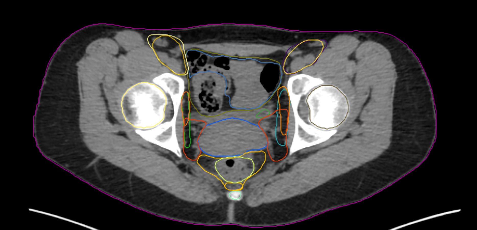
  • Female Pelvis

    Efficient OAR segmentation for CT-based pelvic workflows

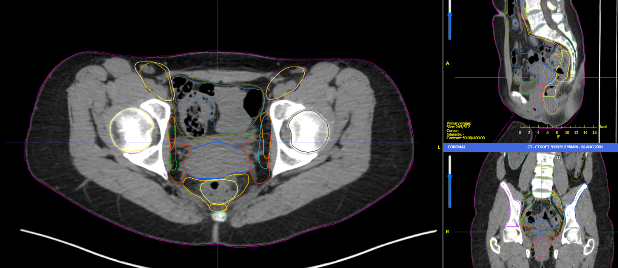
  • Female Pelvis

    Efficient OAR segmentation for CT-based pelvic workflows

  • Brachytherapy (Female)

    MRI-guided OAR delineation for precise brachytherapy delivery

  • Brachytherapy (Female)

    MRI-guided OAR delineation for precise brachytherapy delivery

Prof. Eric Deutsch
Prof. Eric Deutsch

Chair, Dept Radiation Oncology Gustave Roussy Cancer Campus

With ART-Plan we have seen a 5-to-6-fold reduction of the time required for the delineation of organs at risk.

Book a Demo

Optimize Planning

ART-Plan's AI-driven automation delivers high-quality, vendor-agnostic treatment plans in minutes. Eliminating manual variability and accelerating RT planning workflows, ART-Plan helps clinicians spend more time focused on complex cases.

Accurate

Helps to enhance precision, optimize target coverage, and reduce toxicities to healthy tissues.

Scalable

Faster, smarter and reliable - ensures the best possible care for every patient. With high quality routine plans.

Efficient

Expert-level RT plans in just a few minutes - eliminating manual variability and accelerating planning workflows.

Expert-level RT plans in minutes.
Ready for physician review.

Planning

Key Features

AI-powered automatic treatment planning
High-quality plan generation within minutes
Optimized target coverage & OAR sparing
Reduces variability among planners
Clinically validated to ensure comparable and better performance vs manual planning
Automatic export to the TPS
Supports the future of ART workflow

*Note that not all models are available in all markets

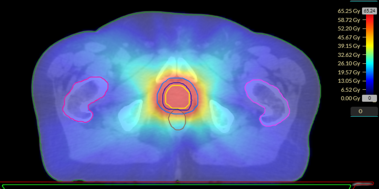
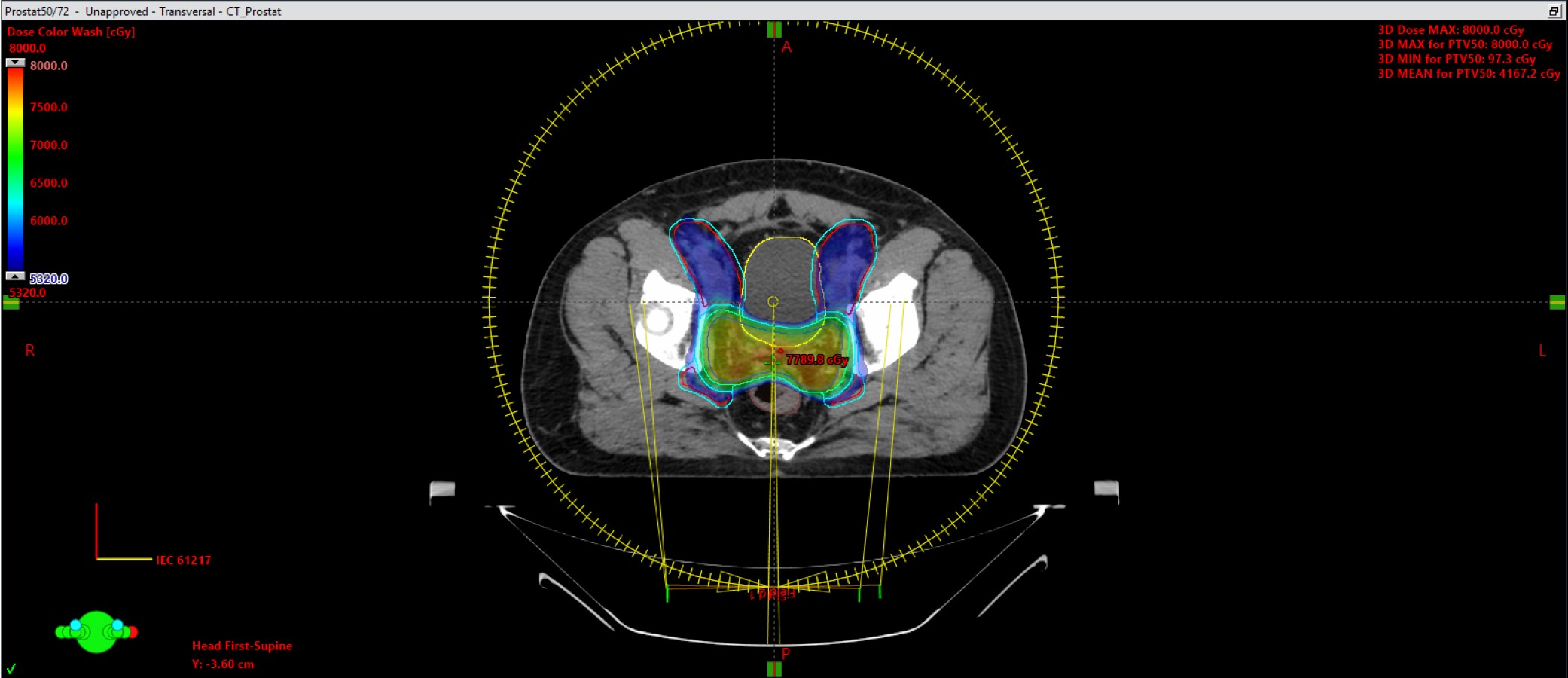
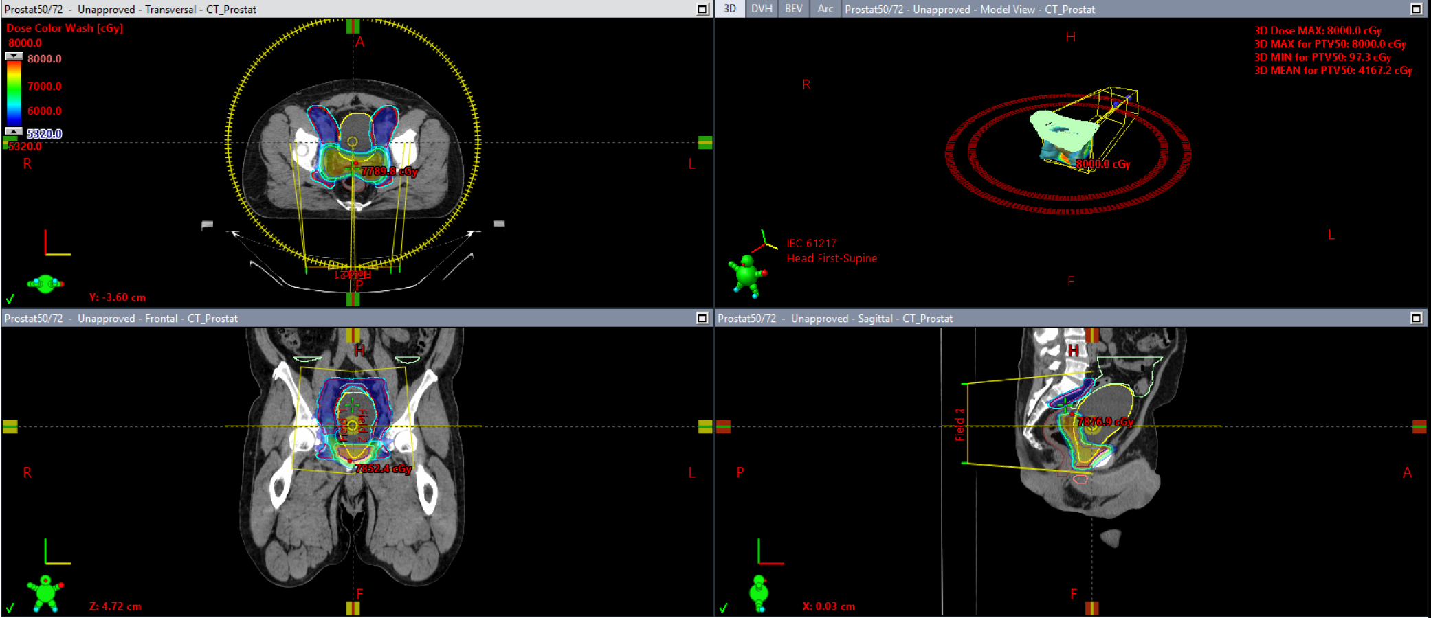
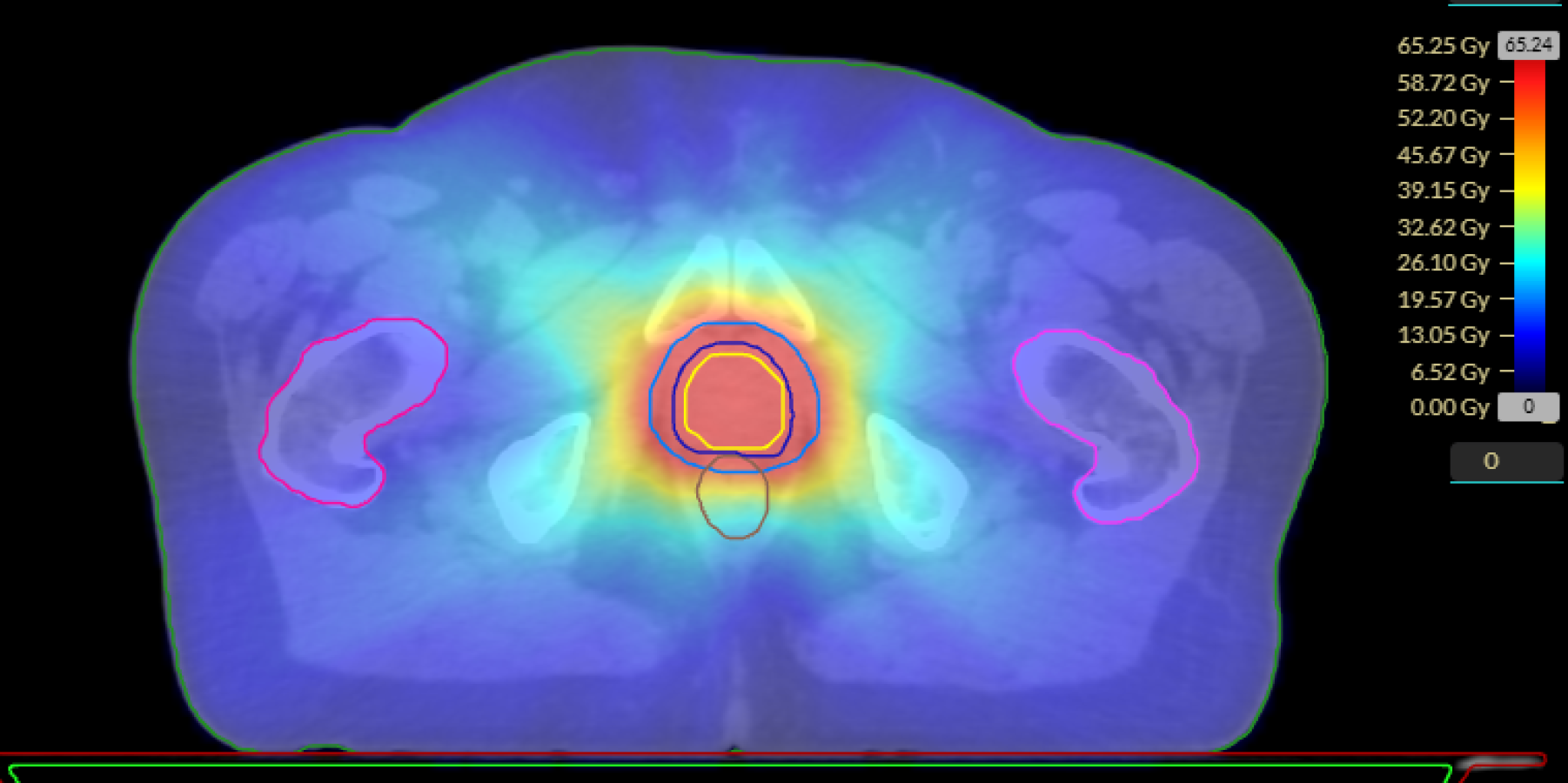
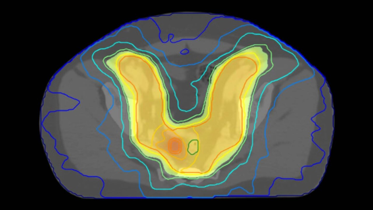
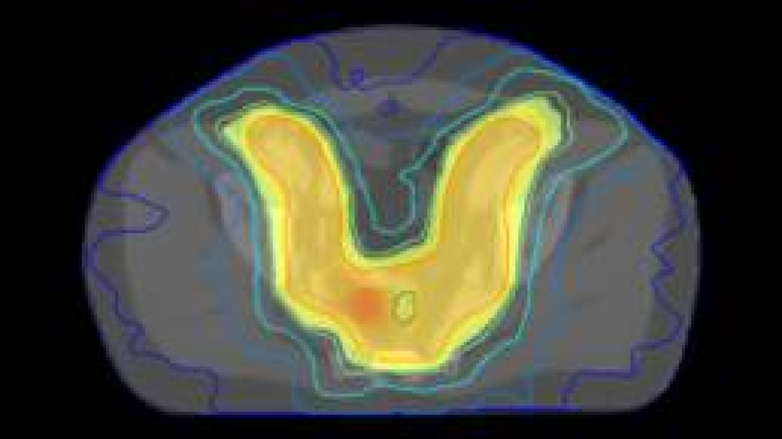
  • High-quality, personalized prostate treatment plans across multiple prescriptions—automated, standardized, and ready in minutes.

  • Your treatment plan is ready—simply calculate the dose in your TPS.

  • Multiple prostate prescriptions. AI-powered planning with consistent clinical quality.

Dr. Alexander Lin
Dr. Alexander Lin

Professor of Radiation Oncology, Penn Medicine

As a ‘one-click’ output solution, SmartPlan demonstrates significant potential for speeding up the treatment planning process while ensuring the best possible treatment for the patient.

Book a Demo

Automate Adaptive

ART-Plan eliminates hours of replanning by automatically reviewing all patient assessments in the background — helping teams make consistent replanning decisions while reducing manual rework.

Consistent

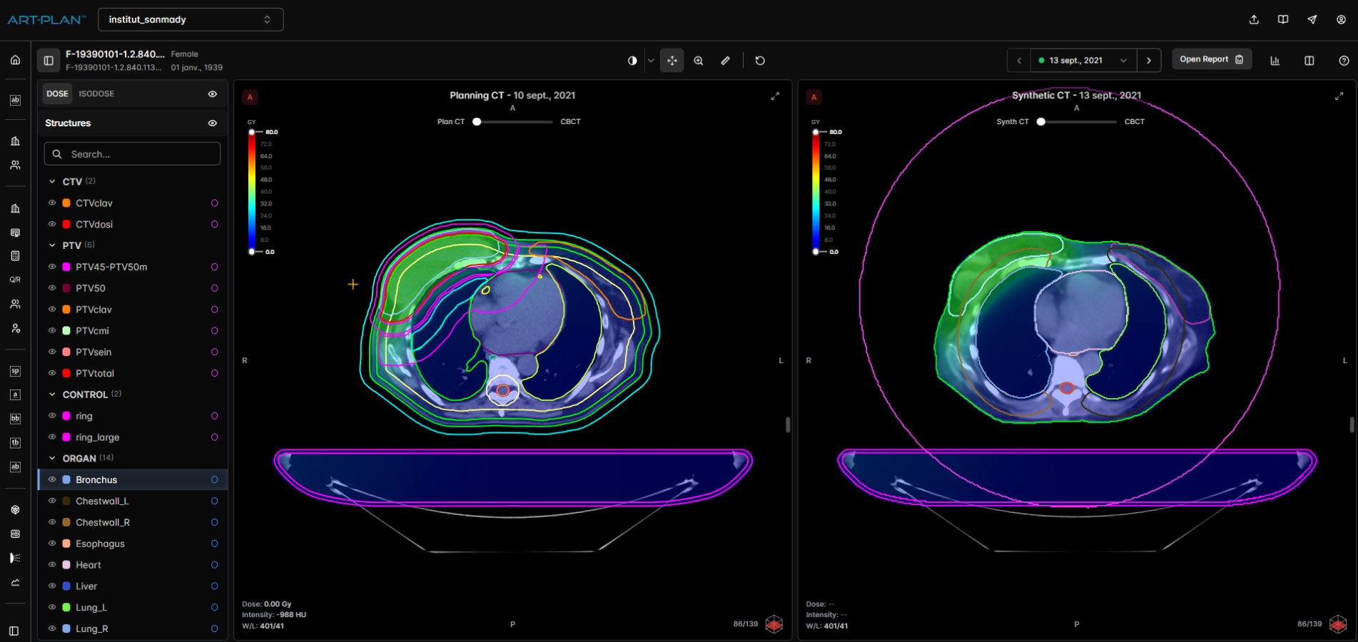
High-quality results through AI generated sCT images, AI DIR and protocol driven metrics that reduce inter-operator variability between assessments.

Seamless

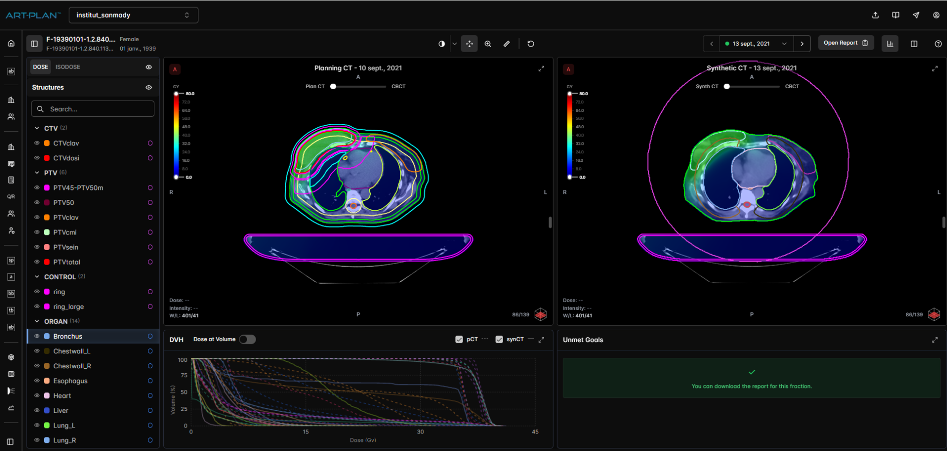
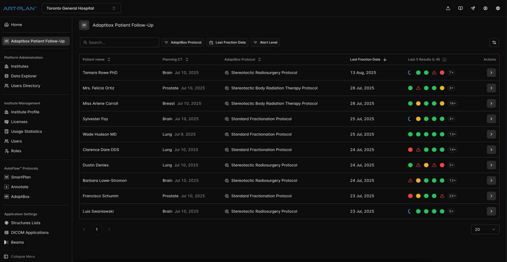
ART-Plan runs in the background to automatically compare CBCT with planning CT of all patients and identifies cases in an intuitive, actionable, and customizable dashboard.

Impactful

Strategic impact beyond adaptive replanning assessment. ART-Plan's data driven approach allows you to focus on high-impact adaptive strategies that inform patient care.

Simplify, Automate and
Implement Adaptive Strategies

Adaptive

Key Features

Seamless interoperability with OIS & TPS
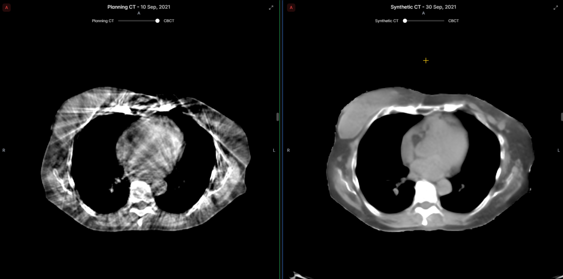
Deep Learning sCT and augmented CBCT
Auto-delineation of OARs
Automated DIR of the planning CT with the CBCT
Propagation of target contours
Automated dose calculation and comparison
Protocol-based alert generation
Customizable dashboard & report templates

*Note that not all models are available in all markets

Adaptive Models

Structures
Library

OARs - Guidelines: OAR as in Gay et al 2012, Mir et al 2020 

Anal_Canal, Bladder, Femur_Head_(L/R), Penile_Bulb, Rectum, Sigmoid_Colon  

Regions of interest 

Prostate, Seminal_Vesicle 

OARs - Guidelines: Kong 2011 (RTOG), Jabbour et al 2014 (RTOG), and Mir et al 2020 

Breast_(L/R), Bronchial_Tree, Chestwall_(L/R), Esophagus, Heart, Lung_(L/R), Spinal_Canal, Spinal_Cord, Trachea, Syn-CT 

OARs - Guidelines: DAHANCA 2020, Eekers et al 2021 (EPTN) and Mir et al 2020 (International consensus) 

Brachial_Plex_(L/R), Brainstem, Esophagus, Eyeball_(L/R), Glottic_Larynx, Larynx, Lens_Eye_(L/R), Lips, Lung_(L/R), Mandible, Optic_Nerve (L/R), Optic_Chiasm, Oral_Cavity, Parotid_(L/R), Pharynx_Const_Muscle, Spinal_Canal, Spinal_Cord, Supraglottic_Larynx, TM_Joint_(L/R) 

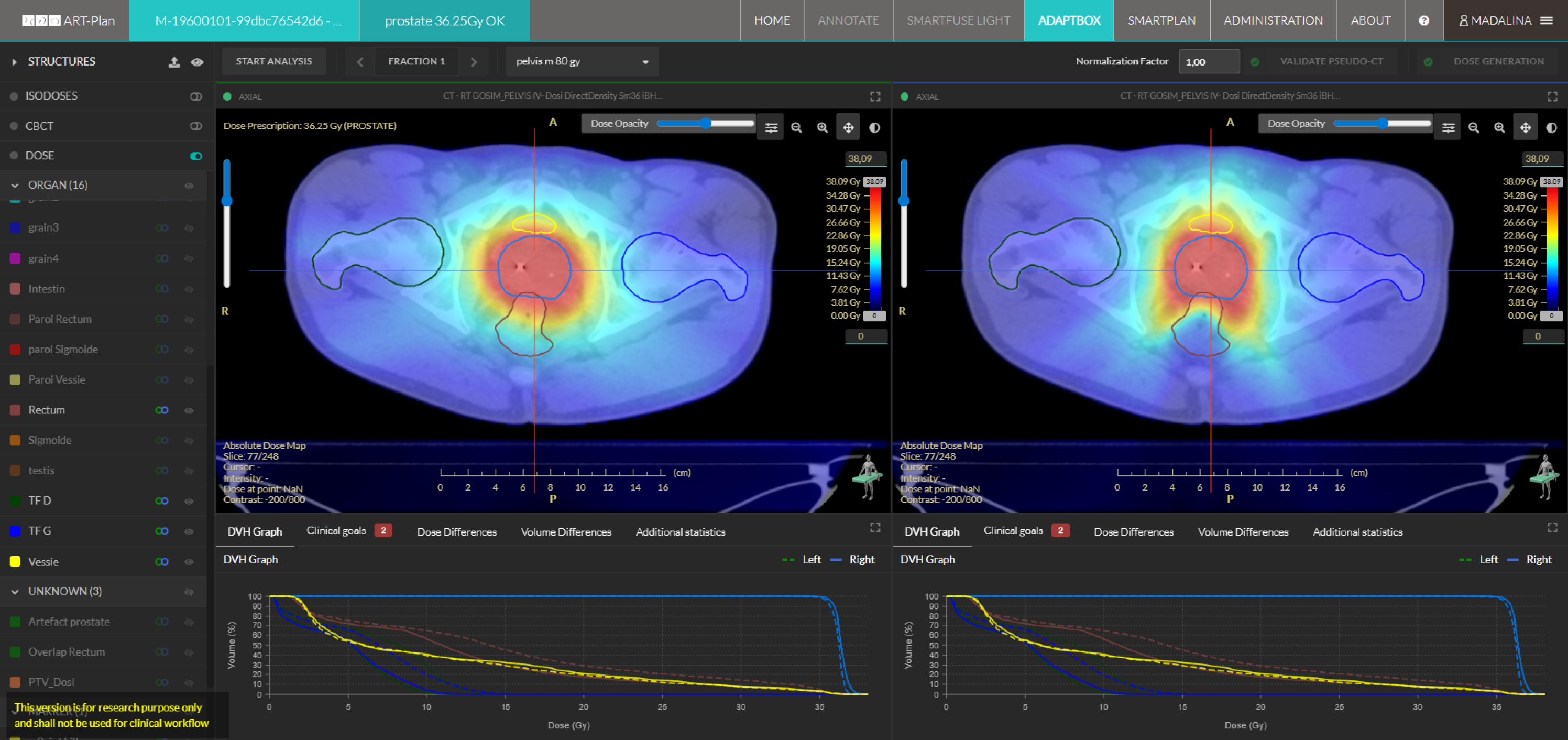
  • Transform daily CBCT into actionable adaptive insights with AI-driven support.

  • Keep every treatment on track with AI-driven quality and decision support.

  • From CBCT to synthetic CT—AI-powered imaging for efficient adaptive radiotherapy.

  • Automated alerts for confident replanning decisions.

Angelique Ducteil
Angelique Ducteil

MD - Radiation Oncologist COPB - Bayonne, France

With AdaptBox's synthetic CT, we validated dose coverage precisely and safely 3 times during treatment, without interrupting care or exposing the patient to additional scans. It saved our team around 3 hours of work - eliminating the need for rescanning, recontouring, replanning, and QA.

Book a Demo